Cathet. Cardiovasc. Diagn. 43: 421-427, 1998
Usefulness of Collagen Plugging with VasoSeal® after PTCA
as Compared to Manual Compression with Identical Sheath Dwell
Times
Sigmund Silber, MD, Aina Björvik, Holger Mühling, MD, Andreas Rösch, MD
Dr. Müller Hospital, Munich, Germany
- This study investigated the usefulness of collagen plugging with VasoSeal® in patients after PTCA compared to a control group having identical sheath dwell times and therefore comparable levels of anticoagulation. A total of 150 patients were enrolled in this prospective and randomized study. Sheaths were pulled at exactly 5 hours after arterial puncture. Time to hemostasis and local complications were determined. There were no statistical differences in baseline characteristics. The mean time to hemostasis in the collagen group was significantly shorter (3 ± 3 minutes) than that of the control group (17.4 ± 7 minutes). At 24 hours, 23% of the collagen group patients had a small, 1% a medium and 4% a large hematoma. In the control group, 32% had a small, 4% a medium sized but no patient a large hematoma. After collagen, one patient developed a pseudoaneurysm needing vascular surgery. In the control group, no major complication occurred.
Compared to patients with manual compression at an identical sheath dwell time and an identical level of anticoagulation, there was a significant reduction in time to hemostasis but no statistical difference regarding local complications. Although the incidence of medium or large hematoma was low, the trend towards a decreased risk of smaller hematomas seemed to be counterbalanced by an increased risk of larger hematomas.
- Key Words: hemostasis, complication, anticoagulation, coronary
artery disease
- INTRODUCTION:
Several randomized trials clearly established that collagen plugging of arterial puncture sites significantly reduces time to hemostasis [1-4]. On the other hand, there are controversial reports whether collagen plugging concomitantly reduces the rate of local complications: Some authors found a significant reduction in local complications [2, 5], whereas other findings were nonconclusive [1]. In contrast, some reported an increased incidence of minor [3] or major complications [4] and therefore described collagen plugging of arterial puncture sites as a "deep disappointment" [6].- In these trials, the collagen groups were not compared to
the control groups at identical conditions: the sheaths were
pulled immediately in the collagen groups, whereas in the control
groups the sheaths were pulled several hours later [2, 5, 7,
8] or even the next day [7, 9]. The sheath dwell time, however,
represents one of the risk factors for local complications [3,
10] and intrinsically determines the level of anticoagulation
and therefore time to hemostasis. Since there is no study comparing
the patients after collagen plugging with a control group at
identical conditions, we performed such a prospective randomized
study with both groups having identical sheath dwell times.
- METHODS:
PTCA's were performed using 8 F sheaths. All patients received 10,000 units of heparin as a standard dose. A reversal of the effect of heparin was not performed. Prolonged duration PTCA patients requiring an additional bolus of heparin, patients with need for overnight heparin infusion or coumadin were not enrolled. Further exclusion criteria were as follows: inadvertent penetration of the dorsal arterial wall with the puncture needle, previous application of collagen sealing of the femoral access site, known allergy to collagen, peripheral artery disease, patients with acute myocardial infarction, status post thrombolytic therapy, known coagulation defects or known platelet dysfunction, severe, uncontrolled arterial hypertension (systolic > 220 mmHg or diastolic > 120 mmHg), preexisting hematoma, hematoma developed during the procedure or patients with a venous femoral sheath. Marked obesity or age were not exclusion criteria, since they do not seem to be related to the rate of vascular complications nor to the success of collagen application [9, 11].
After informed consent was obtained, 150 patients were randomly assigned to either collagen sealing or manual compression. Exactly 5 hours after femoral puncture, sheaths were pulled. Blood pressure and PTT were measured prior to sheath removal. The sheaths were removed independent of these results (normal upper value for PTT in our laboratory is 45 s). In control patients, the sheath was removed and hemostasis obtained with standard manual compression (no C-clamp or other mechanical devices were used). Patients assigned to collagen received local anesthesia before the collagen plug was inserted.
The Vascular Hemostatic Device (VHD, VasoSeal®; Datascope Corp., Montvale, NJ, USA) consists of purified collagen plugs that induce the formation of a hemostatic cap directly over the arterial puncture site. The method of its application is described in detail elsewhere [1, 12]. In brief, VHD comprises four parts: a blunt-tipped 11F dilator, one of seven differently sized 11.5 F sheaths selected by length using a preprocedure needle depth measurement technique and two 90 mg collagen cartridges. When the sheath is pulled, a short guide wire was inserted and the existing sheath was removed while maintaining manual compression. Then the blunt-tipped 11F dilator was inserted over the guide wire just down to the site of the arterial puncture. Guidance was obtained by feeling the resistance of the dilator against the outer surface of the artery as well as by the marker on the dilator. Then the 11.5 F sheath was advanced over the dilator down to the arterial surface. While still holding pressure, the dilator and the guide wire were removed and the collagen cartridge was deployed with a "push and pull" movement. In this study, we used only one cartridge of collagen, since we know from previous studies that one collagen plug is as effective as two plugs but is better tolerated [13, 14]. After the application of the collagen plug, light pressure was sustained for 2 minutes to obtain hemostasis.
By partially releasing manual pressure in the collagen group at 2 minutes after sheath removal, time to hemostasis was measured at 5 minutes, continuing in 5-minute intervals until complete hemostasis was obtained. In the manual group, 5-minute intervals were applied. The protocol for determination of time to hemostasis in the control group was identical. The first time at which no bleeding occurred was taken as the time to hemostasis. Then a pressure dressing was applied to all patients. Since time to ambulation was not an endpoint of this study, patients had to stay in bed until next morning. Patients were then asked to characterize the local symptoms as comfortable, slightly uncomfortable or uncomfortable. Hematoma size was measured just prior to ambulation and described as small (< 7 cm), medium (7 cm to 15 cm) or large (> 15 cm).
According to the VHD multicenter trial [1], major complications were defined as one of the following: thrombosis or loss of distal pulses, large pseudoaneurysm or AV-fistula, bleeding with need for transfusion or any vascular surgery. Bleeding from puncture site not needing transfusion and/or vascular surgery as well as small pseudo-aneurysm treated medically were classified as a minor complication.
Statistics:
Data with normal distribution were analyzed using the two-tailed t-test, variables not normally distributed were analyzed using the unpaired Mann-Whitney U test. For comparison of discrete variables, the chi-square test was used. Results are presented as mean value ± SD; p < 0.05 was considered significant.
VHD:
Control:
patients n = 74
n = 76
female (%) 22
24
age (years) 59.8 ± 9.0
58.0 ± 9.2
mean height (cm) 171 ± 8.5
171 ± 8.7
mean weight (kg) 81.1 ± 11.6
80 ± 12.2
risk factors: smokers 35 %
33 %
hypertension 55 %
59 %
high cholesterol 74 %
71 %
diabetes 25 %
19 %
at sheath removal: systolic BP (mmHg) 125.7 ± 13.6
127.9 ±18.5
diastolic BP (mmHg) 77.8 ± 8.7
75.7 ± 12.1
PTT (s) 49.4 ± 31.0
45.4 ± 33.0
- Table I. Baseline characteristics of enrolled patients
- * There was no statistical difference in demographic characteristics as well as in blood pressure or PTT at time of sheath removal.
RESULTS:
150 patients were enrolled. 74 were assigned to collagen, 76 to the manual control group (Table 1). There were no statistical differences in gender, age, height, weight or cardiovascular risk factors between the two groups (Table 1). Blood pressures and PTTs just prior to sheath removal were also comparable (Table 1).
55 patients had a normal and 19 patients a prolonged PTT in the collagen group, whereas 56 patients of the control group had a normal and 20 a prolonged PTT (figures 1a and 1b). The need for atropine due to vagal reactions during sheath removal was not statistically different between the two groups (0.33 ± 0.58 ml per patient in the collagen group, 0.44± 0.67 ml in the control group).
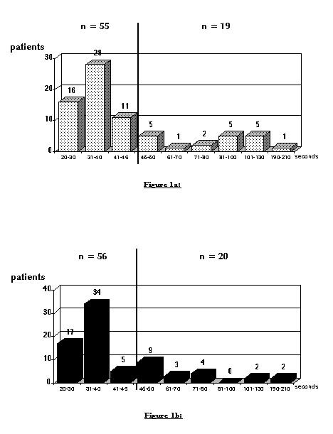
Figure 1: Distribution of PTT's at sheath removal in the collagen group (upper panel) and distribution of PTT's in the control group (lower panel). the vertical line represents the upper limit of normal.
Time to hemostasis:
The mean time to hemostasis in the collagen group was 3 ± 3 minutes (2 - 15 minutes) and statistically shorter (p < 0.001) than that of the control group with 17.4 ± 7 minutes (5 - 75 minutes) (figure 2). At the first check (2 minutes), 63 patients with VHD showed complete hemostasis, yielding a hemostasis success rate of 85% at 2 minutes (figure 2).
The answers to the inquiries the next morning regarding the patients' classification of their groin discomfort revealed "comfortable" in 90.5% of VHD and 97% of control patients, "slightly uncomfortable" in 6.7 % of VHD and 3 % of control patients. "Uncomfortable" was answered in 2.8 % of patients receiving VHD. There was no statistical difference between both groups.
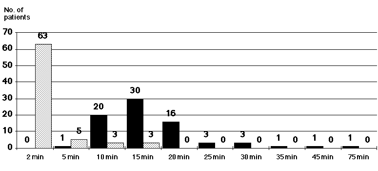
Figure 2: Presence of complete hemostasis at each checking interval in patients that received VHD (dotted bars) compared to the control group (black bars).
Local Complications:
Major local complications: one patient, after collagen application and rapid hemostasis (2 minutes, PTT at time of insertion was 68 s), developed a large hematoma; ultra-sound revealed a pseudoaneurysm needing vascular surgery. In the control group, no major complication occurred.
Minor local complications: At 24 hours after sheath pulling 17 collagen group patients (23%) had a small, one patient (1%) a medium and 3 patients (4%) a large hematoma. In the control group, 24 patients (32%) had a small, 3 (4%) a medium sized but no patient a large hematoma (figure 3). There was no statistical difference between the groups regarding the development of a hematoma.
The mean duration of hospital stay was 4.1 ± 4.2 days in the collagen group and 5.2 ± 6.9 days in the control group (not significant).
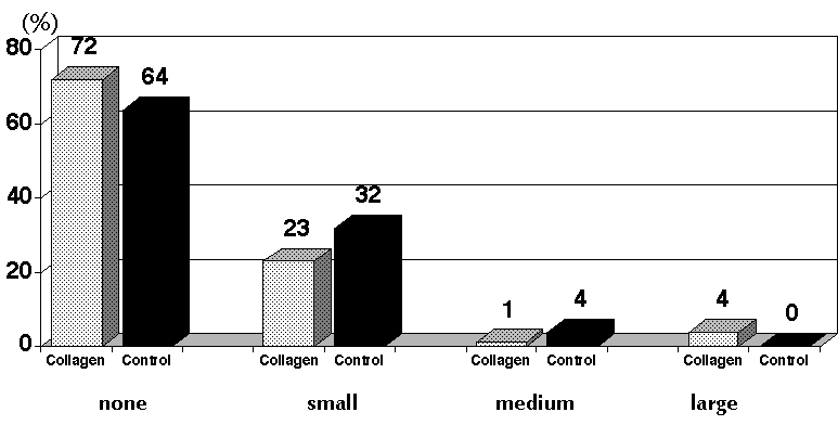
Figure 3: Presence and size of hematomas in the collagen (dotted bars) and control (black bars) groups. Data are displayed in percent of patients.
DISCUSSION:
After PTCA, a sheath dwell time of > 24 hours is a risk factor for local complications [10]. Another risk factor is prolonged anticoagulation [10, 11]. Since PTCA patients in whom sheaths are left indwelling overnight require prolonged anticoagulation, either one of these risk factors could lead to an increased incidence of bleeding [3, 11]. Therefore, the reported advantage of a sealing device in reducing local complications may have been artifactual due to an increased risk of hemorrhage in the control group caused by longer sheath dwell times [2, 5]. On the other hand, a control group with immediate sheath removal and manual compression would have been unethical in PTCA patients [1]. Only one study reported a collagen group with late sheath removal, but no data was provided regarding the sheath dwell times [1].
As our results show, collagen sealing of femoral arterial puncture sites in patients after PTCA at comparable PTT's and identical sheath dwell times significantly reduces time to hemostasis, but does not reduce groin complications.
Purified bovine collagen has been used in vascular, abdominal and dental surgical procedures since late 1960 as an adjunct to hemostasis when control of bleeding by ligature or other conventional methods was insufficient [15-17]. The biodegradable collagen plug induces local platelet activation and aggregation, releasing coagulation factors and resulting in the formation of fibrin and the subsequent generation of a thrombus [18]. It is assumed that anticoagulation with heparin or even antiaggregation with aspirin does not affect hemostasis induced by collagen [12, 19]. Collagen is ultimately degraded and resorbed by granulocytes and macrophages. These cells, releasing their collagenase enzymes, invade the plug and selectively digest the collagen as a function of the physical properties of the different collagens [20]. Antigenicity of purified collagen is considerably reduced and, although allergies to collagen are described [21], allergic reactions to VHD have not been a clinical problem [1, 2, 12].
Time to hemostasis:
Time to hemostasis is defined as the time elapsed from initial compression at removal of the sheath until the completion of compression. However, the measurement of time to hemostasis is not standardized: results for time to hemostasis intrinsically depend on the time interval to the first and between the subsequent checks for bleeding. Of course, the choice of the time interval between the bleeding checks is an ambiguous decision: too short intervals may not be sufficient to give enough time for thrombus formation and increase - particularly in the manual control groups - the time to hemostasis. In three studies, all patients automatically received a pressure dressing [22], vascular C-clamp [23] or an air cushion device [7], so that the determination of time to hemostasis was not possible.
In our study we decided on a rather short time interval of 2 minutes for the first check, with subsequent measurements at 5 minute intervals, in order to make both groups as comparable as possible. Comparing our results for time to hemostasis in PTCA patients to those of others is limited to only three studies (Table 2). Our somewhat lower numbers for time to hemostasis in the VHD group (3 minutes vs. 5 to 7 minutes) and also in our control group (17 minutes vs. 27 to 33 minutes) is explained by the study design regarding the time of sheath removal and the corresponding level of anticoagulation (Table 2). Nevertheless, our findings regarding the significant reduction of time to hemostasis are in accordance with the other studies (Table 2).
| Authors |
No. of Pat. |
Randomized to |
Sheath Removal |
ACT(s) or PTT(s) |
Time to Hemostasis (min) |
| Sanborn et al. [1] |
n= 85 |
VHD-on heparin |
immediate |
52.9 ± 50.9 |
7.6 ± 11.6* |
|
n= 71 |
VHD-off heparin |
delayed |
36.2 ± 16.9 |
4.3 ± 3.7* |
|
|
n=134 |
control |
delayed |
37.9 ± 18.2 |
33.6 ± 24.2 |
|
| Slaughter et al. [3] |
n= 51 |
VHD |
immediate |
381 ± 152 |
5* (3-15) |
|
n= 50 |
control |
delayed |
151 ± 71 |
27 (18-40) |
|
| v. Hoch et al. [4] |
n=117 |
VHD |
immediate |
- |
5* (4-6) |
|
n=114 |
control |
2-3 hrs |
- |
27 (20-32) |
|
| current study |
n= 74 |
VHD |
5 hrs |
49.4 ± 31.0 |
3 ± 3* (2-15) |
|
n= 76 |
control |
5 hrs |
45.4 ± 33.1 |
17.4 ± 7 (5-75) |
Table II: Published data on time to hemostasis using VHD as compared to control groups in randomized studies in PTCA patients.
Differences may be explained based on different study designs regarding time of sheath removal and levels of anticoagulation. * = significant difference (p < 0.05) as compared to control.
Local Complications:
The complication rates reported for collagen plugging are somewhat confusing, because several studies did not differentiate between diagnostic and interventional procedures and various classifications of complications with different methods of measurement were used: In the European multicenter trial, only the overall complications were reported for the 105 patients receiving the collagen plug after diagnostic and 140 patients after interventional procedure, despite significant differences in the doses of heparin (5715 ± 4615 U vs. 15378 ± 3025 U). ACTs or PTTs were not reported [12].
In our study, a learning curve was not relevant due to our prior extensive experience with this sealing device [14]. According to the classification used [1], one major complication occurred in the collagen group in our study and none in the control group. When comparing minor complications in both groups, no statistical difference was found for the development of any hematoma. Combining patients with small and medium sized hematoma into one group, however, results in statistically significant differences: 18/74 patients assigned to collagen significantly less often developed a small/medium sized hematoma than 27/76 patients assigned to manual compression (24% vs. 36%, p < 0.05). In contrast, significantly more patients assigned to collagen (4%) developed a larger hematoma than patients assigned to conventional sheath pulling (0%). There is an overall trend towards reduction of minor hematomas, counterbalanced by an increased risk of larger hematomas or even a major complication. This may in part explain some of the controversial findings mentioned above.
LIMITATIONS:
The fact that patients assigned to collagen did not feel more comfortable than patients after manual hemostasis may be related to our study design. Using VasoSeal® for sheath pulling immediately after PTCA, patients' comfort may be different [1-3]. Furthermore, our study was not addressed to prove whether length of hospital stay may be reduced by VHD. The decision to discharge was left to the physicians on the wards. Nevertheless, patients with collagen sealing were discharged one day earlier than the control group. This is in good agreement with a US study, which reported a significant decrease from 2.4 ± 0.98 days (control group, 56 patients) to 1.53 ± 0.8 days (collagen group, 47 patients) [25]. The overall length of stay in our study (4 vs. 5 days) seems rather long and may in part be explained by the compensation system of the German health insurance companies (pay by day and not by a fixed fee for the whole procedure). To prove the possible cost reduction of a reduced hospital length of stay would probably require a large-scale trial [26].
Power calculations:
Whereas the VHD-related reduction in time to hemostasis from 17.4 to 3 minutes is highly significant and clinically relevant, the statistical power regarding local complications in our study is limited. Assuming a rate of major local complications in a control group of 2% and a device-related reduction to 1%, one would need to perform a randomized study (alpha-error of 0.05, 2-sided) in 46,590 patients to achieve a power of 80 % and in 60,984 patients for a power of 90% . These data simply do not exist for any hemostatic device. A recent overview of all relevant publications regarding collagen devices [27] and the meta-analysis of the published randomized studies in 6007 patients [28] could not detect a device-related reduction in major local complications.
CONCLUSIONS:
The use of a single collagen plug in patients after PTCA is highly effective for achieving rapid hemostasis. As compared to patients with manual compression at an identical sheath dwell time and therefore at an identical level of anticoagulation, there is no statistical difference regarding local complications. Although the incidence of medium or large hematoma was low, the trend towards decreasing smaller hematomas is counterbalanced by an increased risk of larger hematomas or a major complication and may in part explain some of the published controversial findings.
References:
1, Sanborn TA, Gibbs HH, Brinker JA, Knopf WA, Kosinski EJ, Roubin GS: A multicenter randomized trial comparing a percutaneous collagen hemostasis device with conventional manual compression after diagnostic angiography and angioplasty. J Am Coll Cardiol 22:1273-1279,1993.
2, Schräder R, Steinbacher S, Burger W, Kadel C, Vallbracht C, Kaltenbach M: Collagen application for sealing of arterial puncture sites in comparison to pressure dressing: a randomized trial.Cathet Cardiovasc Diagn 27:298-302, 1992.
3, Slaughter PM, Chetty R, Flintoft VF, Lewis S, Sykora K, Beattie DM, Schwartz L: A single center randomized trial assessing use of a vascular hemostasis device vs. conventional manual compression following PTCA: what are the potential resource savings? Cathet Cardiovasc Diagn 34:210-214,1995.
4, v. Hoch F, Neumann F-J ,Theiss W , Kastrati A, Schömig A: Efficacy and safety of collagen implants for haemostasis of the vascular access site after coronary balloon angioplasty and coronary stent implantation. Europ Heart J 16:640-646, 1995.
5, Nagtegaal EM, Schalij MJ, Buis B: Routine use of collagen to seal the femoral artery puncture site after percutaneous transluminal coronary angioplasty in fully anticoagulated patients; a clinical evaluation. J Am Coll Cardiol 21: 231A, 1993.
6, Geschwind HJ: Percutaneous arterial approach revisited. Eur Heart J 16:579-580, 1995.
7, Camenzind E, Grossholz M, Urban P, Dorsaz PA, Didier D, Meier B: Collagen application versus manual compression: a prospective randomized trial for arterial puncture site closure after coronary angioplasty. J Am Coll Cardiol 24:655-662, 1994.
8, Legrand V, Doneux P, Tilman Chu S: Comparison of puncture site closure with collagen plug (Vasoseal) or by early manual compression following PTCA. Circulation 88:I-72, 1993.
9, Foran JPM, Patel D, Brookes J, Wainwright: Early mobilisation after percutaneous cardiac catheterisation using collagen plug (VasoSeal) haemostasis. Br Heart J 69:424-429, 1993.
10, Muller DWM, Shamir KJ, Ellis SG, Topol EJ: Peripheral vascular complications after conventional and complex percutaneous coronary interventional procedures. Am J Cardiol 69:63-68, 1992.
11, Bogart DB, Bogart MA, Miller JT, Farrar MW, Barr WK, Montgomery MA: Femoral artery catheterization complications: a study of 503 consecutive patients. Cathet Cardiovasc Diagn 34:8-13, 1995.
12, Ernst SMPG, Tjonjoegin RM, Schräder R, Kaltenbach M, Sigwart U, Sanborn TA, Plokker HWT: Immediate sealing of arterial puncture sites after cardiac catheterization and coronary angioplasty using a biodegradable collagen plug: results of an international registry. J Am Coll Cardiol 21:851-855, 1993.
13, Silber S, Haentsch C, Seidel N, Mühling H: Early ambulation (30 minutes) after cardiac catheterization using the collagen plug. Efficacy and follow-up comparing two different plug dosages in 660 cases. J Am Coll Cardiol 21:150A, 1993.
14, Silber S, Dörr R, Rösch A, Mühling H: Complications after collagen plugging for hemostasis following femoral puncture: experience in over 3000 patients. Circulation 92:I-56, 1995.
15, Abbott WM, Austen WG: The effectiveness and mechanism of collagen-induced topical hemostasis. Surgery 78:723-729, 1975.
16, Peacock E, Siegler H, Biggers P: Use of tanned collagen sponges in the treatment of liver injuries. An Surg 161:238-243, 1965.
17, Silverstein ME, Chvapil M: Experimental and clinical experiences with collagen fleece as a hemostatic agent. J Trauma 21:388-393, 1981.
18, Chvapil M, Holubec H: Effect of hemostatic fleece on 14C-Serotonin release by human platelets. Jpn Pharmacol Ther 18:57(2913)-62(2918), 1990.
19, Chvapil M, Chvapil TA: Hemostatic effectiveness of hemostatic collagen fleece (Novacol) in heparinized and Aspirin treated rabbits. Jpn Pharmacol Ther 18:43(2899)-48(2904), 1990.
20, Chvapil M: Tissue reaction and biodegradation of implanted hemostatic collagen fleece in rats. Jpn Pharmacol Ther 18:179 (3927)-192(3940), 1990.
21, Kitamura K, Yasuoka R, Ohara M, Shimotsuma M, Hagiwara A, Yamane T, Yamaguchi T, Takahashi T: How safe are the xenogeneic hemostats? Report of a case of severe systemic allergic reaction. Surg Today 25:433-435, 1995.
22, Kiemenej F, Laarman GJ: Next-day discharge after transradial Palmatz-Schatz coronary stenting: a first step towards outpatient coronary stent implantation. Circulation 90:I-620, 1994.
23, Webb JG, Carere RA, Dodek AA: Collagen plug hemostatic closure of femoral arterial puncture sites following implantation of intracoronary stents. Cathet Cardiovasc Diagn 30:314-316, 1993.
24, Bartorelli AL, Sganzerla P, Fabbiocchi F, Montorsi P, De Cesare N, Child M, Tavasci E, Passaretti B, Loaldi A: Prompt and safe femoral hemostasis with a collagen device after intracoronary implantation of Palmaz-Schatz stents. Am Heart J 130:26-32, 1995.
25, Spokojny AM, Fahey F, Gibbs HH, Molloy T, Shaftel PA, Barra L, Sanborn TA: Use of collagen plug immediately post angioplasty in heparinized patients allows earlier discharge. Circulation 88:I-252, 1993.
26, Krause PB, Klein LW: Utility of a percutaneous collagen hemostasis device: to plug or not to plug? J Am Coll Cardiol 22:1280-1282, 1993.
27, Silber S: Rapid Hemostasis of Arterial Puncture Sites with Collagen in Patients Undergoing Diagnostic and Interventional Cardiac Catheterization. Clin Cardiol 20:981-992, 1997.
28, Silber S: Hemostasis success rates and local complications with collagen after femoral access for cardiac catheterization: Analysis of 6007 published patients. Am Heart J 135:152-156, 1998.
- Correspondence to:
- Sigmund Silber, MD, FACC
- Professor of Medicine
- Dr. Müller Hospital
- Am Isarkanal 36
- D-81379 München
- Tel: (+ 49 89) 74 21 51-0
- Fax (+ 49 89) 74 21 51 31
- e-mail: ssilber@med.de